--------Ongoing
Studies--------------------

B. Functional structure revealed by
voltage sensitive dye imaging
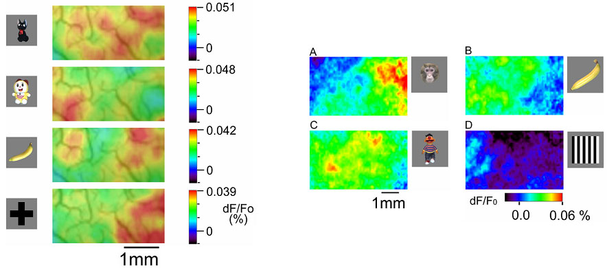
Voltage sensitive dye imaging revealed
that two components of fluorescence changes were elicited by visual stimuli:
local modulation of fluorescence changes (left panel, red region), and spread
of fluorescent changes over one millimeter (right panel, active domain indicated
by green region). The active domains were stimulus specific (right panel).
The local modulation coincided with activity spots revealed by intrinsic
signal imaging. The result suggests the existence of functional domain whose
area was larger than columns.

B. Functional structure revealed by
voltage sensitive dye imaging
Ryota Homma
