Research
- Biomaterials for regenerative medicine
- Nanomedicine
- Molecular diagnosis and imaging
- Nucleic acid and protein engineering
- Nano-materials and nano-interfaces
1.1 Immobilization of growth factors and binding growth factors
In the field of regenerative medicine and tissue engineering, three components such as cells, extracellar matrix and growth factors are important. This laboratory has been working on combination of growth factors and the matrix (materials). Not only chemical immobilization of growth factors on material surfaces, but also protein engineering was used for the synthesis of binding growth factors and applied in animal experiments.
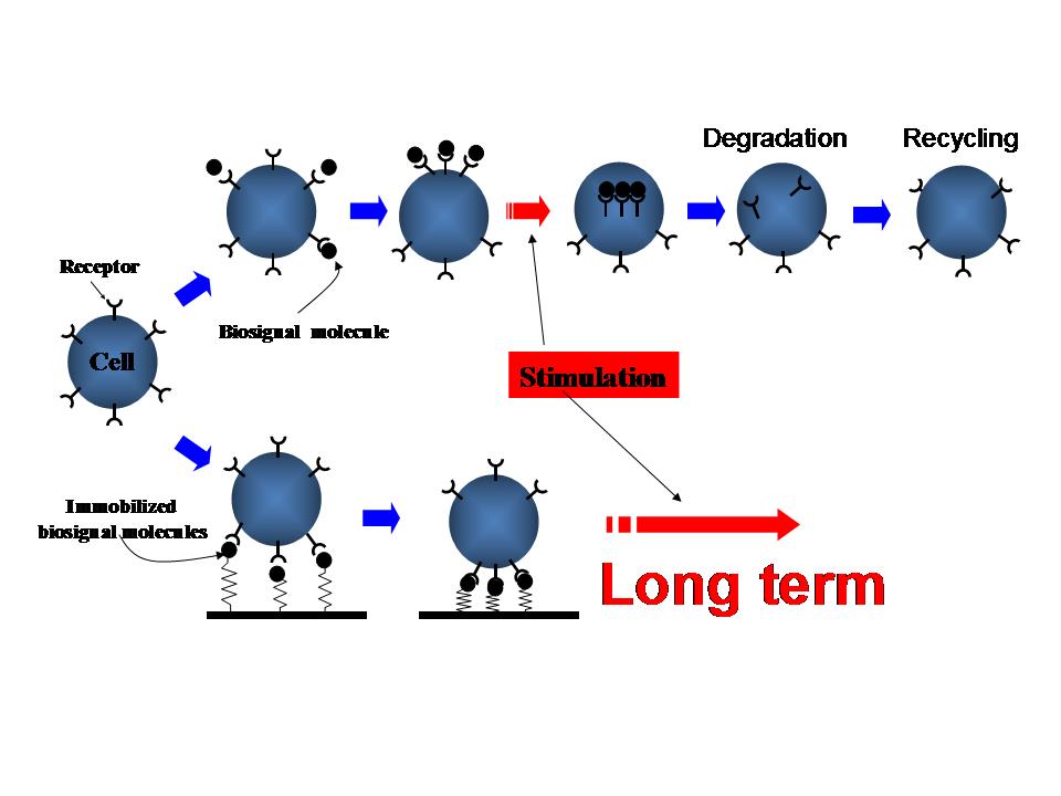 Figure 1 Long-term activation by immobilized biosignal molecule
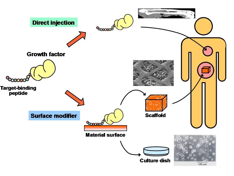 Figure 2 Applications of engineered growth factors
1.2 Photo-reactive biomaterials
UV-curable biopolymers were prepared from synthetic polymers and biological macromolecules including chitosan, recombinant human gelatin, fish gelatin, and hyaluronic acid. They were employed for micropatterning of proteins and visualize the effect of immobilized proteins. Visible light curable ones were also prepared from biological macromolecules including gelatin and chitosan and they were employed as bio-adhesives or bio-sealants.
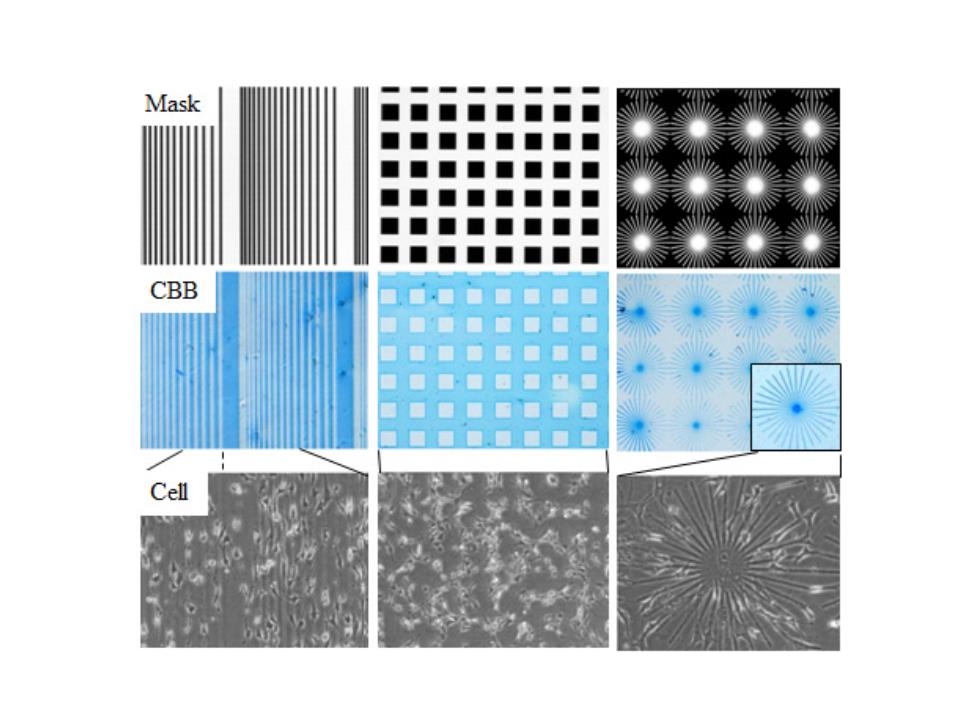 Figure 3 Micropatterned mask (upper), micropattern immobilizations of protein and staining with CBB (middle), and cell culture on micropatterned surfaces (lower)
1.3 ES and iPS cell culture
Our first target was the development of biomaterials for ex vivo expansion of stem cell was cord blood cells. For this purpose a telomerase gene was incorporated into human stromal cells and the immortalized cells were used as a nurse cell for expansion of hematopoietic stem cells. The second target was expansion of artificial stem cells involving mouse, monkey embryonic stem (ES) cells, and iPS cells. For this purpose, chemically fixed feeder cells and artificial materials including protein-immobilized surfaces were developed.
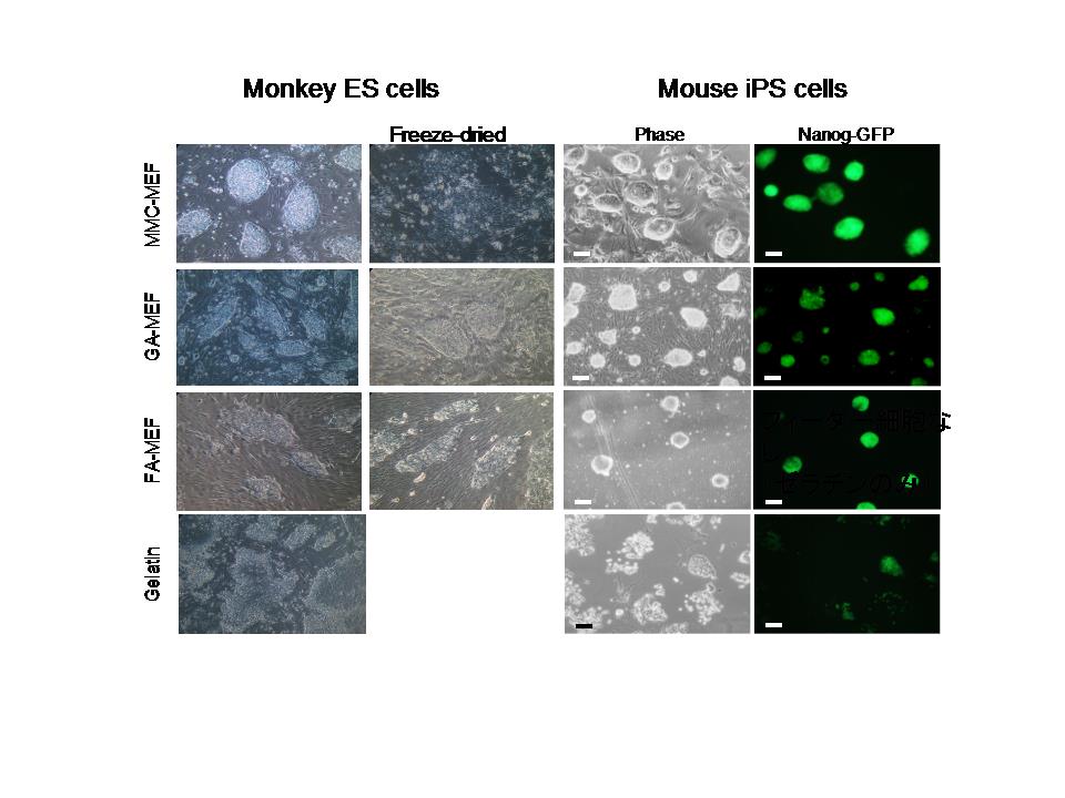 Figure 4 Culture of monkey ES and mouse iPS cells on mitomycine C-treated mouse embryonic feeder (MMC-MEF), glutaraldehyde-fixed MEF (GA-MEF), formaldehyde-fixed MEF (FA-MEF), and gelatin.
1.4 Cell manipulation by MEMS
In order to prepare stem cells derived from somatic cells of which the method is called initialization or reprograming, some methods have been employed as illustrated in Figure 1-24. We have been developing a new method which uses cell fusion with an embryonic stem cell to avoid the nuclear transfer or iPS method by the gene transfection.
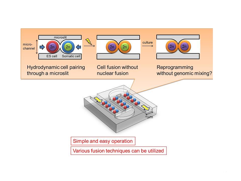 Figure 5 Cell manipulations by Micro Electro Mechanical Systems (MEMS)
2.1 Design of siRNA (nanostructured siRNA)
RNA interference, in which short interfering RNA (siRNA) molecules are used to target specific genes for down regulation in vivo, is among the most powerful molecular biology techniques to emerge in recent years. Many scientists see considerable promise in clinical applications of this technology, although there are a number of serious technical roadblocks that remain to be overcome. We designed and synthesized new structured RNAs for RNA interference applications.
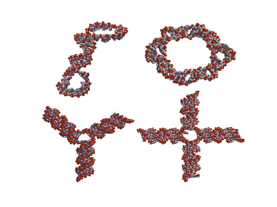 Figure 6 Nano-structured siRNAs
2.2 Drug delivery devices
The importance of drug delivery system and the device for the purpose has significantly increased. Therefore we have devised some new delivery systems for vaccines, siRNA, and medical drugs. Biodegradable biochip was also fabricated for the design of controlled-release drug-delivery systems.
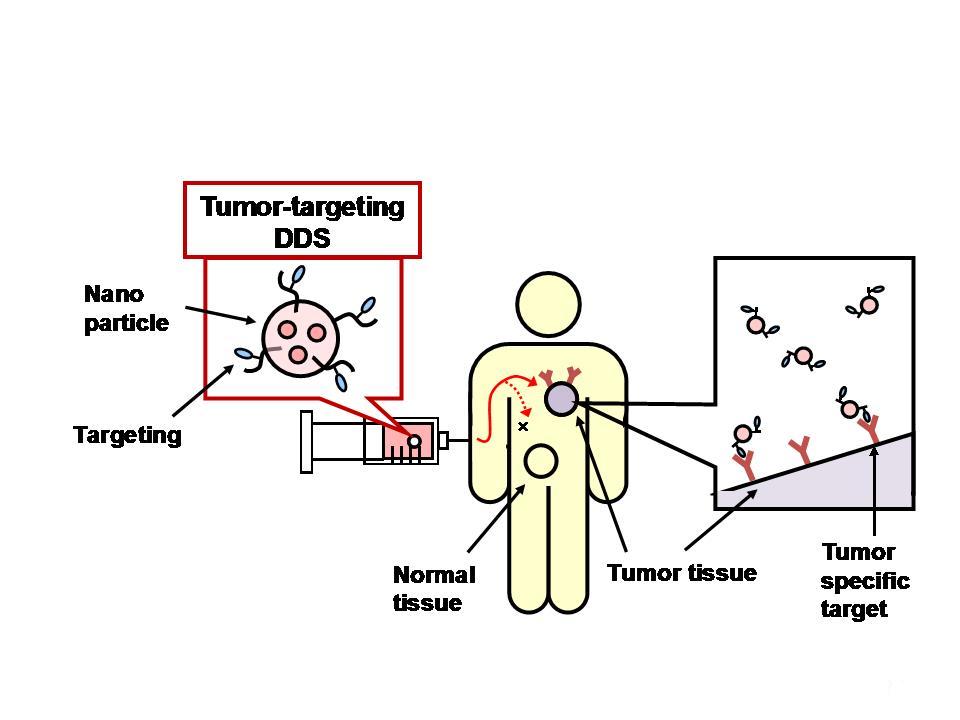 Figure 7 An example of drug delivery system
3.1 Protein Microarray biochip
Microarray technology has become a crucial tool for large-scale and high-throughput biological science and technology. It allows fast, easy, parallel detection of thousands of addressable elements in a single experiment under exactly the same conditions. However, protein microarray technology has not developed as rapidly compared with DNA microarray technology because of the difficulty in immobilizing proteins. We have devised a new method, which is a photoimmobilization technique, for the preparation of microarrays. To date we have used this technique to immobilize proteins, viruses, and cells on a solid matrix to investigate the interaction of biological components and to apply to clinical diagnoses. By using this technology, Tashiro and Ito have set up a business as a RIKEN venture company, Consonal Biotechnologies, Co. Ltd.
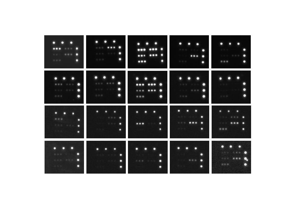 Figure 8 Detection image of microarray chip
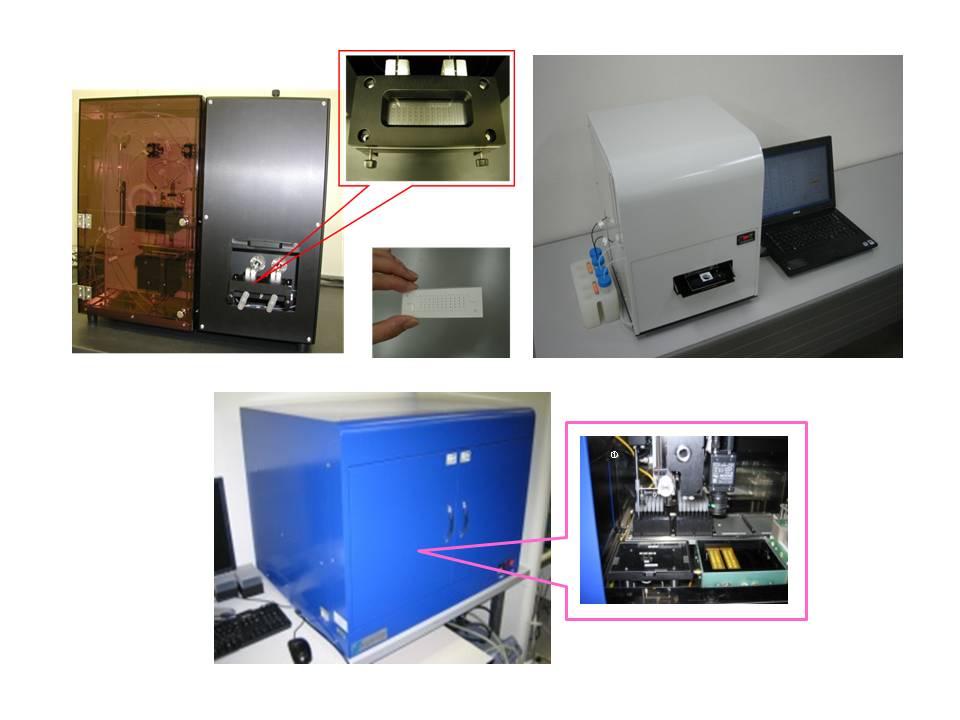 Figure 9 Automated microarray systems
3.2 Molecular probes for detection of genes and proteins
Bioimaging or biomonitoring of living cells is a very important tool for academic research. However, there had been no practical method to detect gene expression in living cells. We have succeeded in the development of a reduction-triggered fluorescence (RETF) probe that shows a high signal/background ratio for sensing oligonucleotides. The probes were activated only by a specific reducing reagent on the oligonucleotide target and were very stable under biological conditions, showing little background fluorescence. Glutathione transferases (GSTs) are used in biotechnology applications as fusion partners for facile purification, known to detoxify endogenous compounds such as peroxidized lipids, and breakdown products of xenobiotics and are overexpressed in tumor cells. We developed a new fluorescent probe for detection of GST. In addition, now we are developing new molecular probes by molecular evolutionary engineering.
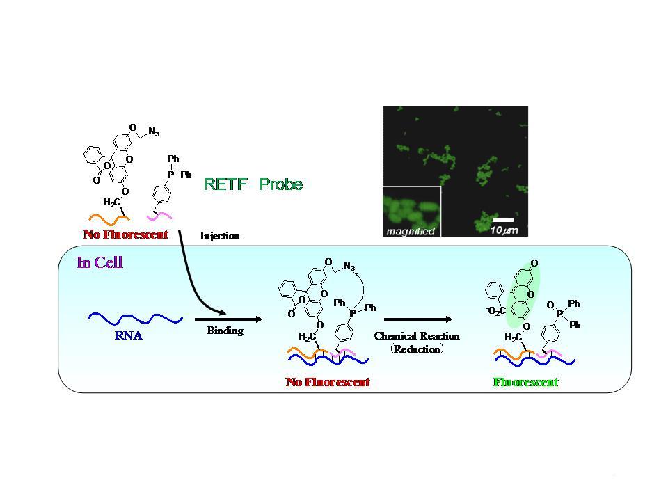 Figure 10 Mechanism of developed RETF probe
4.1 Bioorthogonal protein and oligonucleotide engineering
Bioorthogonal protein synthesis is performed using ribosome during translation process. Genetic PEGylation or limitless protein synthesis using cyclic RNA template is achieved by the ribosome. Antibodies or oligonucleotides were covalently conjugated with PEG and the properties of the PEGylated ones in organic media were investigated.
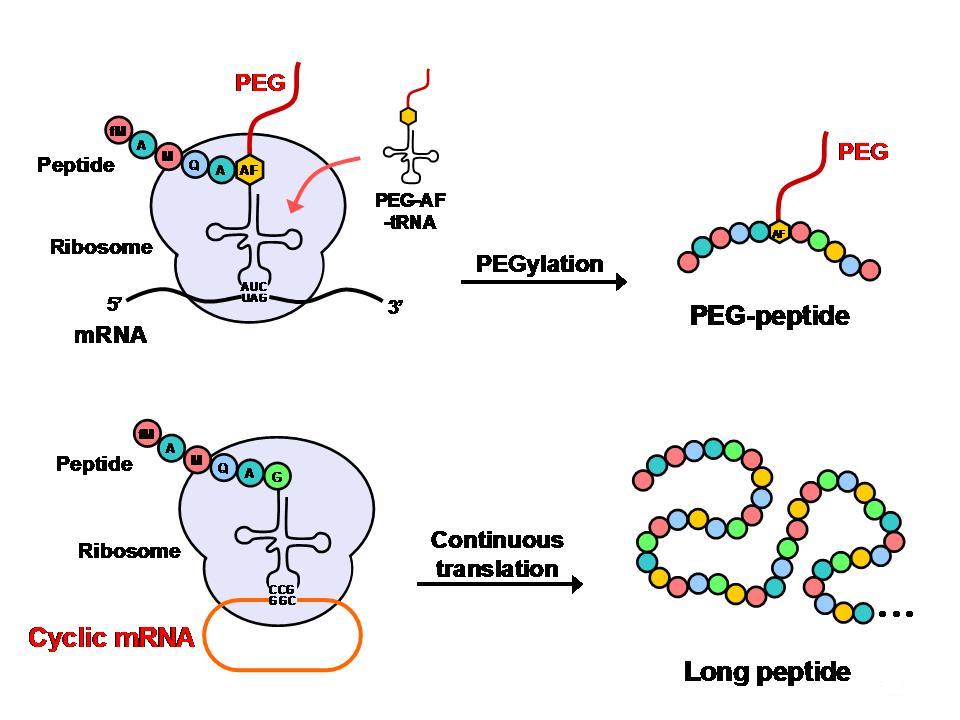 Figure 11 Ribosomal syntheses of bioorthogonal proteins
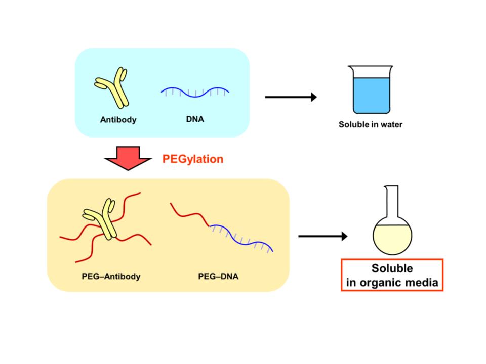 Figure 12 PEGylated antibody and oligonucleotides are active in organic media
4.2 Molecular evolutionary engineering
The selection mimics Darwinian evolution was also called molecular evolutionary engineering. The selected oligonucleotides or peptides are now called as aptamers. The evolutionary engineering opened a new window to evolve aptamers artificially by starting from a library including unnatural oligonucleotides or peptides. Incorporation of unnatural oligonucleotides into a selection library was achieved using enzymatic reactions (PCR or RNA polymerase). On the other hand, for incorporation of unnatural amino acid misacylated tRNA was required. The incorporated number of unnatural components in the selection library is increasing, and we call this transitional period of selection library moving from “natural soup” to “unnatural soup”. In this laboratory first DNAzyme and ribozyme recognizing hemin were in vitro selected. Secondly by using azobenzene-carrying adenine triphosphate in vitro selection to the target of hemin was performed. Now various types of new peptide aptamers have been developed in our laboratory.
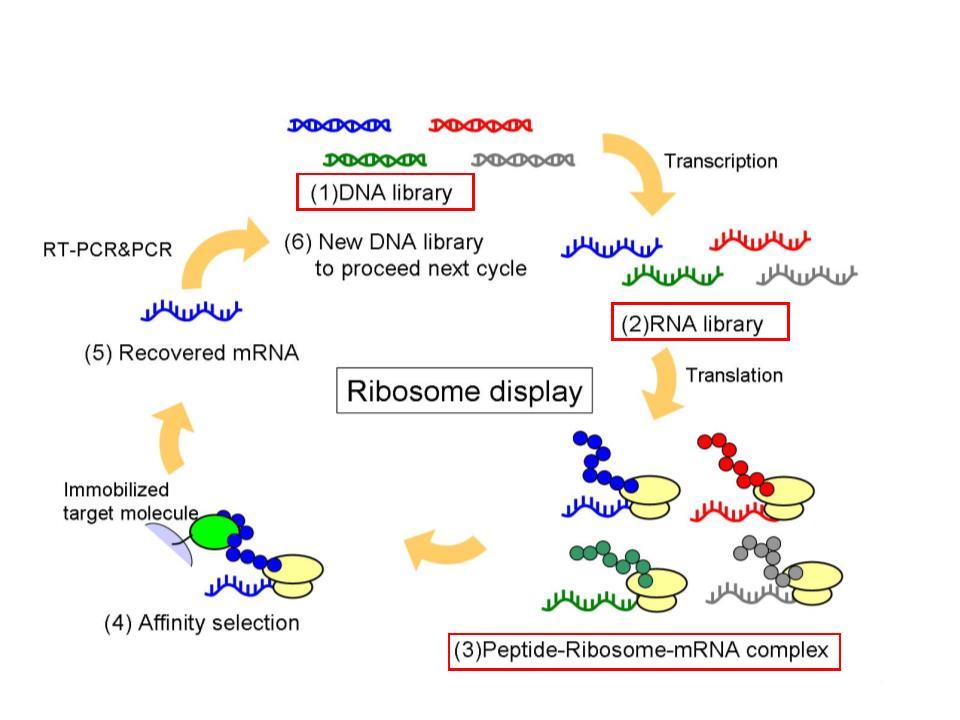 Figure 13 Ribosome display
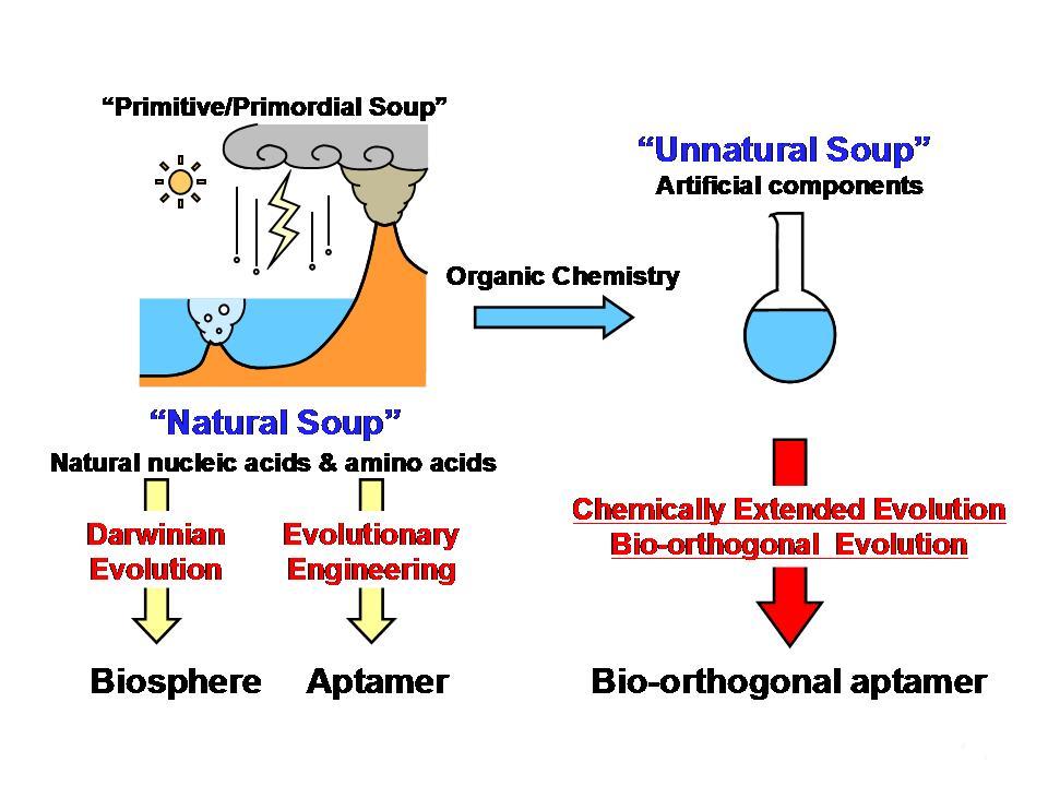 Figure 14 Extension of molecular evolutionary engineering
5.1 Stimuli-responsive polymers
Several new types of stimuli (pH, ionic strength, temperature, photo)-responsive polymers have been synthesized. A thermoresponsive polymer poly(N-isopropylacrylamide) carrying a Ru complex, a catalyst of the Belousov?Zhabotinsky reaction, was synthesized in collaboration with Prof. Yoshida at the University of Tokyo and immobilized on a glass plate. Periodic turbidity changes in the aqueous solution of the polymer were observed, and nanoscale self-oscillation of the immobilized polymer was observed by a scanning probe microscope.
5.2 Surface treatments (Wettability control and non-biofouling polymers)
Surface gradation has been performed by using photolysis or photo-reactive polymers. By copolymerization of vinyl monomers containing zwitterion residues and photo-reactive moiety, several photo-immobilizable non-biofouling polymers were synthesized. Photo-immobilization of the polymers reduced protein adsorption or cell adhesion onto the modified materials.
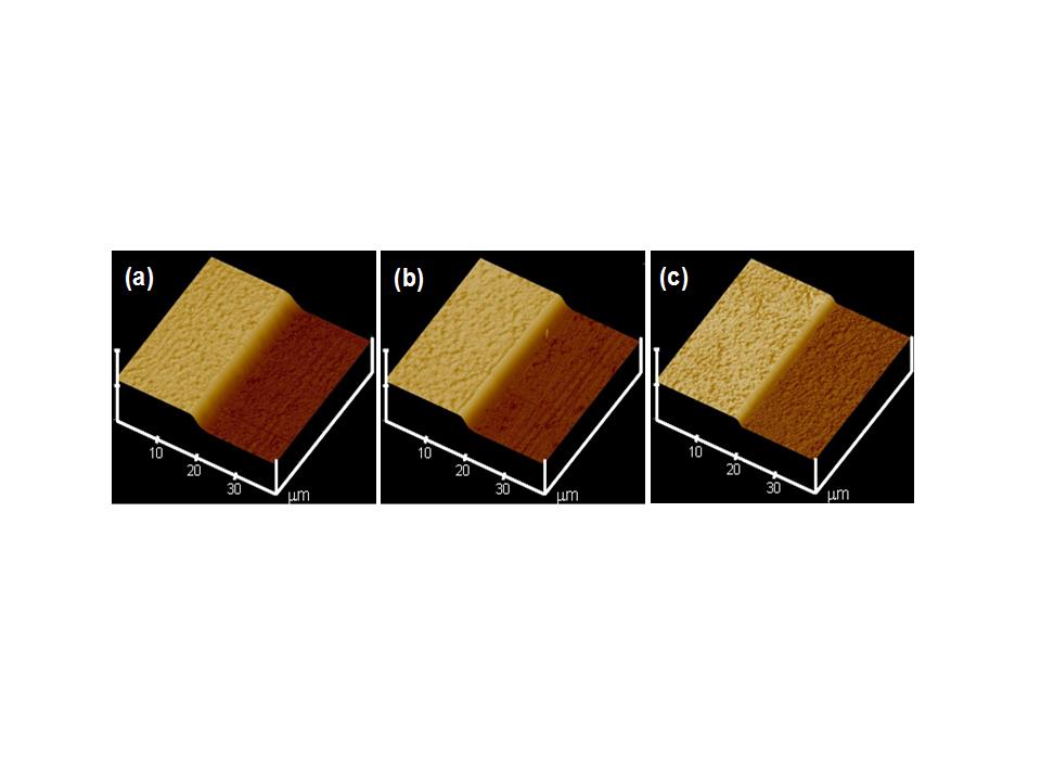 Figure 15 AFM images of micropatterned polyethylene glycol on (a) titanium, (b) glass, and ThermanoxTM
5.3 Nano-fibers by electrospinning method
Biodegradable nano-fibers have been prepared by electrospinning method and the fibers were conjugated with other materials for medical applications.
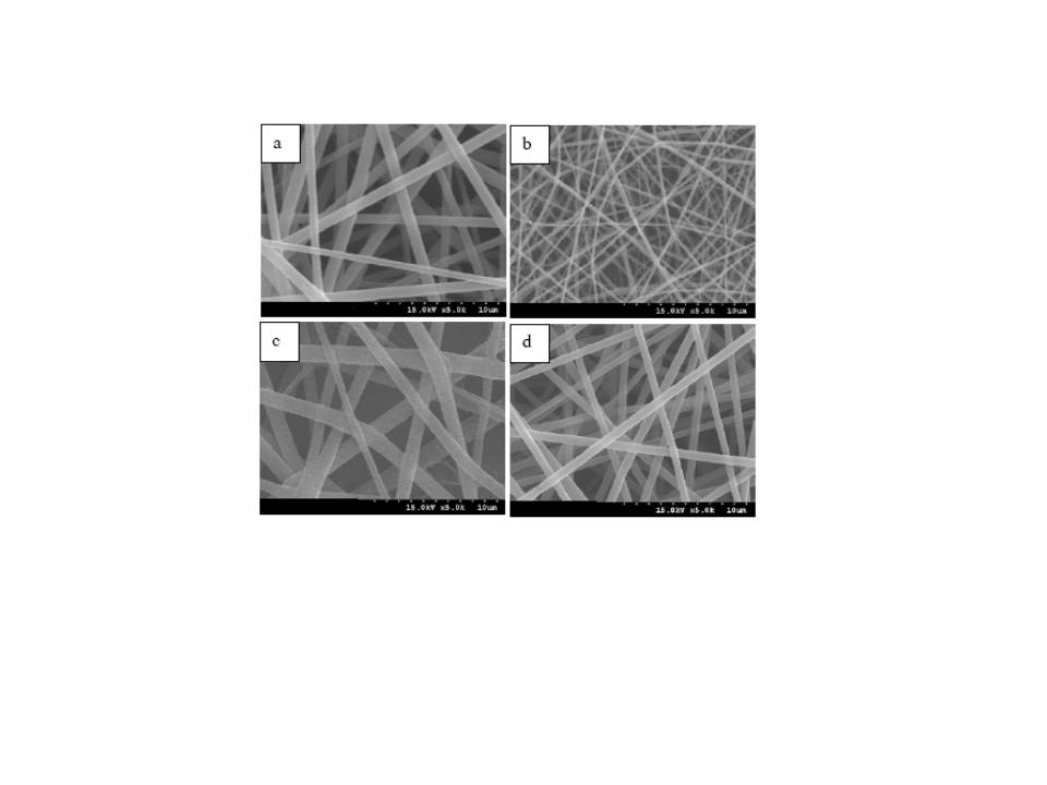 Figure 16 SEM micrographs of electrospun fibers of PHBV/gelatin (50/50) using TFE solutions as a function of concentration. (a) 2 wt%, (b) 4 wt%, (c) 6 wt%, (d) 8 wt%.
